✅ Respiratory Motion Management Series ④ - A Premium Version of CPAP? NIMV60p
📍Introduction
In the previous issue, I introduced CPAP as a technique that uses continuous positive airway pressure to expand the lungs and shift the heart and other normal organs into more favorable positions without requiring the patient to hold their breath.
However, what CPAP raises is the baseline pressure of breathing. Above that baseline, when the patient inhales and exhales, how deeply they breathe, and how regular that breathing remains are still largely up to the patient. As a result, although lung volume increases, CPAP does not necessarily reduce the magnitude or variability of respiratory motion, particularly the movement of tumors located near the diaphragm, in a consistent manner.
So what if we could take this one step further? Could we keep the lungs expanded while also making the patient’s breathing itself faster and shallower?
This is the idea behind NIMV60p.
With NIMV60p, different pressures are applied during inspiration and expiration, similar to bilevel positive airway pressure (BiPAP), while the respiratory rate is increased substantially. This leaves very little time for the patient to insert spontaneous breaths between ventilator-delivered breaths. The patient increasingly follows the rapid, shallow breathing pattern generated by the machine rather than initiating spontaneous breaths of their own.
In this final issue of the series, I would like to introduce NIMV60p, a respiratory motion management technique that combines the lung inflation effect of CPAP with respiratory regularization and motion suppression.
📍Instead of Asking the Patient to Control Their Breathing
One of the most common approaches to maintaining regular breathing during radiation therapy is simply to ask the patient to control their breathing.
We may tell them, “Breathe comfortably and regularly,” or “Try to breathe a little more shallowly,” or use visual and audio guidance to help them follow a predefined breathing cycle.
The problem is that breathing changes as soon as we start thinking about it. It changes when we become nervous or tired, and the longer treatment continues, the more difficult it becomes to maintain the same breathing pattern that was established at the beginning.
In 2016, Parkes and colleagues approached this problem from an entirely different direction. Rather than asking patients to breathe well, they allowed a ventilator to take over the breathing pattern.
Fifteen patients with breast cancer received mechanical ventilation through a face mask while remaining awake, without sedation or anesthesia. It took less than one minute for the ventilator to take control of the breathing pattern, and all patients completed the procedure without discomfort.
Within-patient variability in respiratory rate decreased by 85%, while variability in inflation volume decreased by 29%. Variation in the end-inspiratory and end-expiratory positions of a breast surface marker was only 0.5 mm and 0.4 mm, respectively.
Perhaps the most interesting point was that patients did not need to concentrate on their breathing or watch a screen to keep themselves synchronized.
Instead of the patient controlling the breathing, the machine created a regular breathing pattern for them.
📍From Making Breathing Regular to Making It Smaller
Researchers then began using mechanical ventilation not only to make breathing more regular, but also to reduce the size of each breath itself.
Van Ooteghem and colleagues evaluated several forms of non-invasive mechanical ventilation, or NIMV.
In a volume-controlled mode, respiratory rate and tidal volume were kept constant. In a shallow-controlled mode, respiratory rate was increased while the volume of each breath was reduced. In a slow-controlled mode, high and low pressures were alternated to create repeated mechanically induced DIBH.
In other words, the same ventilator could produce very different respiratory states depending on its settings: regular breathing, rapid shallow breathing, or even repeated breath holds.
Among these approaches, rapid shallow breathing was particularly interesting for thoracic and upper-abdominal tumors affected by respiratory motion.
As respiratory rate increases, the amount of time available for each individual breath becomes shorter. If minute ventilation is maintained, the tidal volume of each breath becomes smaller, which can in turn reduce the excursion of the diaphragm and tumor.
West and colleagues applied rapid shallow ventilation at respiratory rates of 20/min and 25/min to 10 healthy volunteers and measured internal motion using MRI.
Compared with free breathing, internal organ motion decreased by 56% and 62%, respectively. In the posterior right lung, where the largest motion was observed, mean motion decreased from 40.0 mm during free breathing to 15.5 mm and 15.2 mm, respectively.
Simply increasing the respiratory rate resulted in smaller breaths with less variability.
Researchers at Amsterdam UMC also evaluated regularized breathing at a respiratory rate of 22/min.
Among 15 healthy volunteers, median diaphragm motion decreased by 39%, from 20.9 mm during free breathing to 12.8 mm.
However, substantial residual motion remained in some participants. The breathing pattern had become more regular, but at 22 breaths per minute, there was still enough time for patients to insert spontaneous breaths or take breaths with relatively large tidal volumes.
This naturally led to the next question:
“Would increasing the respiratory rate even further reduce motion?”
📍NIMV60: Breathing Once Every Second
Researchers at Amsterdam UMC increased the respiratory rate to 60/min.
That means a complete inspiration and expiration every second.
As with CPAP, positive end-expiratory pressure, or PEEP, was also applied so that positive airway pressure was maintained at the end of expiration and the lungs remained in an expanded state.
The ventilator maintains PEEP during expiration and briefly applies a higher pressure during inspiration.
For example, if PEEP is maintained at 15 cmH₂O and inspiratory pressure is set 15 cmH₂O above PEEP, peak inspiratory pressure reaches approximately 30 cmH₂O.
The difference between inspiratory and expiratory pressure creates each small breath, while the higher PEEP raises the baseline lung volume around which those breaths occur.
Once the respiratory rate reaches 60/min, each respiratory phase becomes extremely short. It becomes difficult for the patient to insert a deep or irregular spontaneous breath between ventilator-delivered breaths, so the patient instead follows the rapid, shallow breathing pattern created by the ventilator.
This approach, in which breathing is not stopped completely but the machine effectively takes control of its rate and depth, can be described as forced rapid shallow breathing.
In the early studies from Amsterdam UMC, the emphasis was on the respiratory rate of 60/min, and the technique was therefore called NIMV60.
Later, to distinguish the current technique from rapid ventilation at RR 60/min without PEEP, the combination of RR 60/min and PEEP became known as NIMV60p. The “p” refers to PEEP.
📍RR 60 Reduces Motion, While PEEP Expands the Lungs
In an early study presented at ESTRO in 2022, six healthy volunteers underwent MRI while receiving ventilation at RR 60/min, with PEEP increased stepwise from 0 to 5, 10, and 15 cmH₂O.
Even without PEEP, simply increasing the respiratory rate to 60/min reduced median diaphragm motion by 76%, from 37 mm during free breathing to 11 mm.
Motion decreased in every participant.
As PEEP increased to 5, 10, and 15 cmH₂O, sagittal lung area increased by 18%, 31%, and 41%, respectively, compared with ventilation without PEEP.
In other words, the technique combined the lung inflation effect provided by CPAP with the motion suppression effect of rapid shallow ventilation.
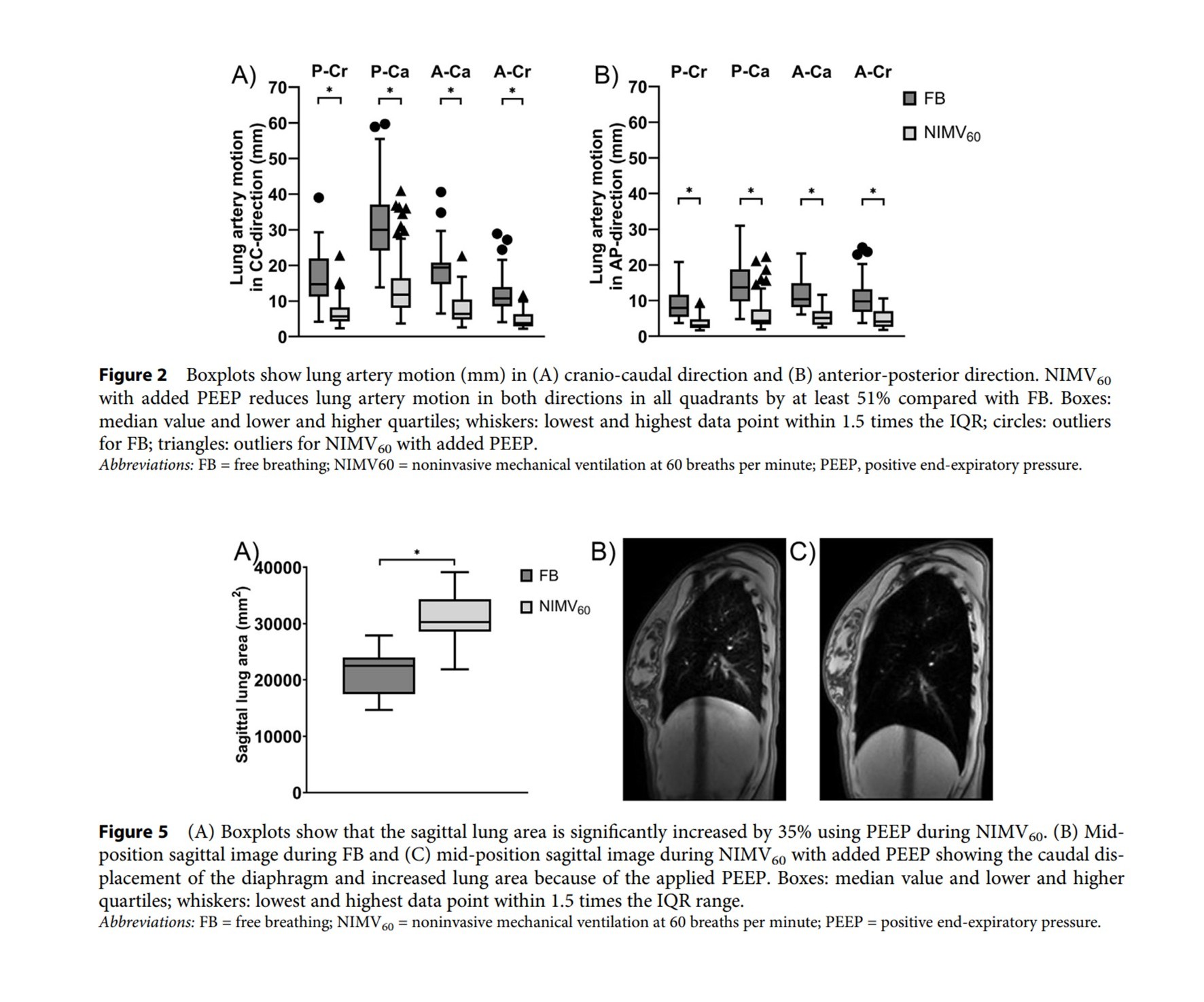
Veldman-Landegent and colleagues later analyzed the effects of NIMV60p in greater detail in a 2025 study involving 12 healthy volunteers.
Using cross-sections of the pulmonary arteries as surrogates for lung tumors, they found that craniocaudal motion decreased by 61–67%, while anteroposterior motion decreased by 51–68%.
Sagittal lung area increased by 35% compared with free breathing, and MRI demonstrated expansion of the lungs as the diaphragm shifted in the caudal direction.
In other words, it was not only the diaphragm that moved less. Motion of structures within the lungs also decreased consistently across multiple locations and directions. |